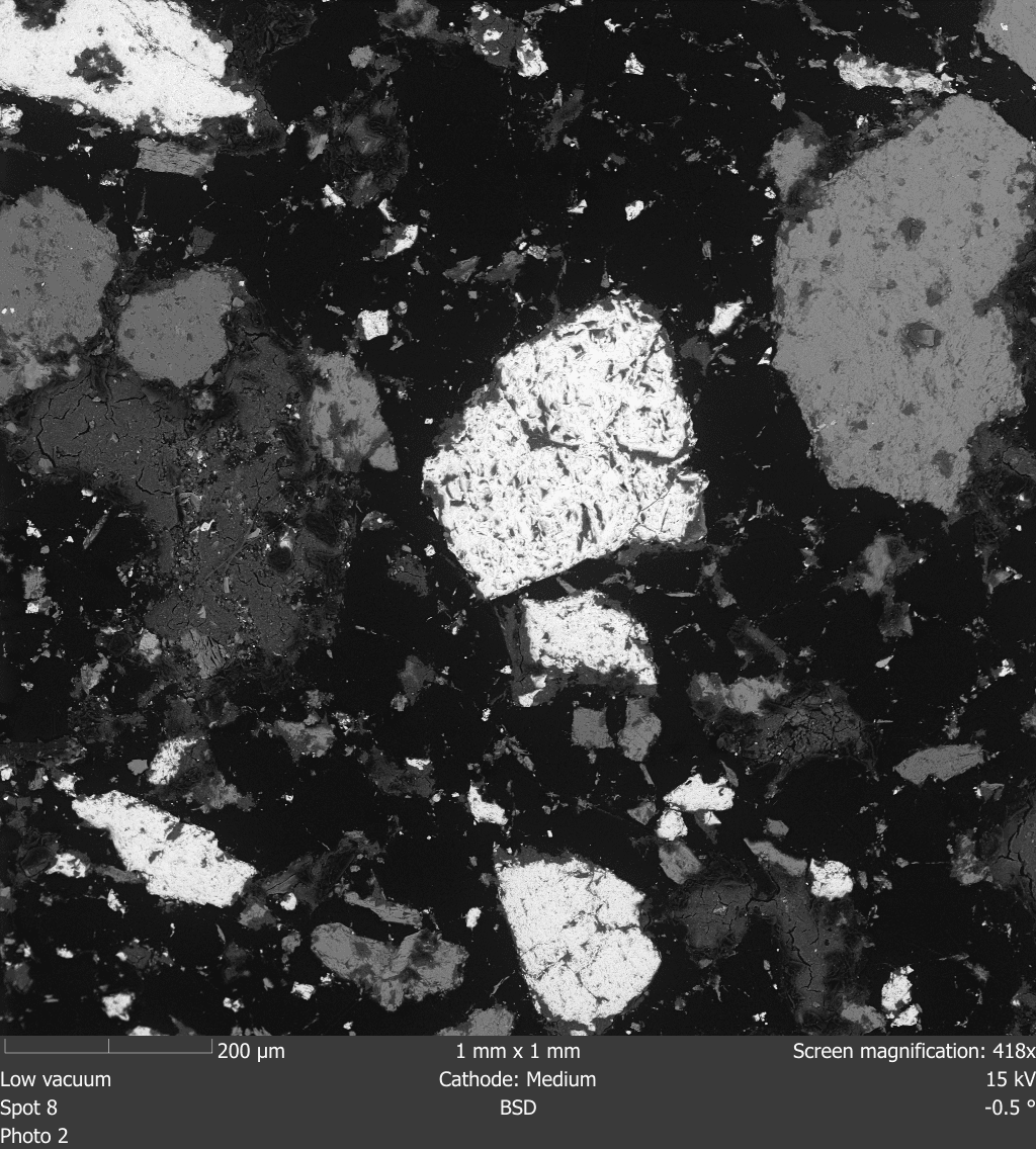
Semplor SEM backscattered-electron image of a polished iron-ore specimen

Fig 01 · SEM image of iron ore

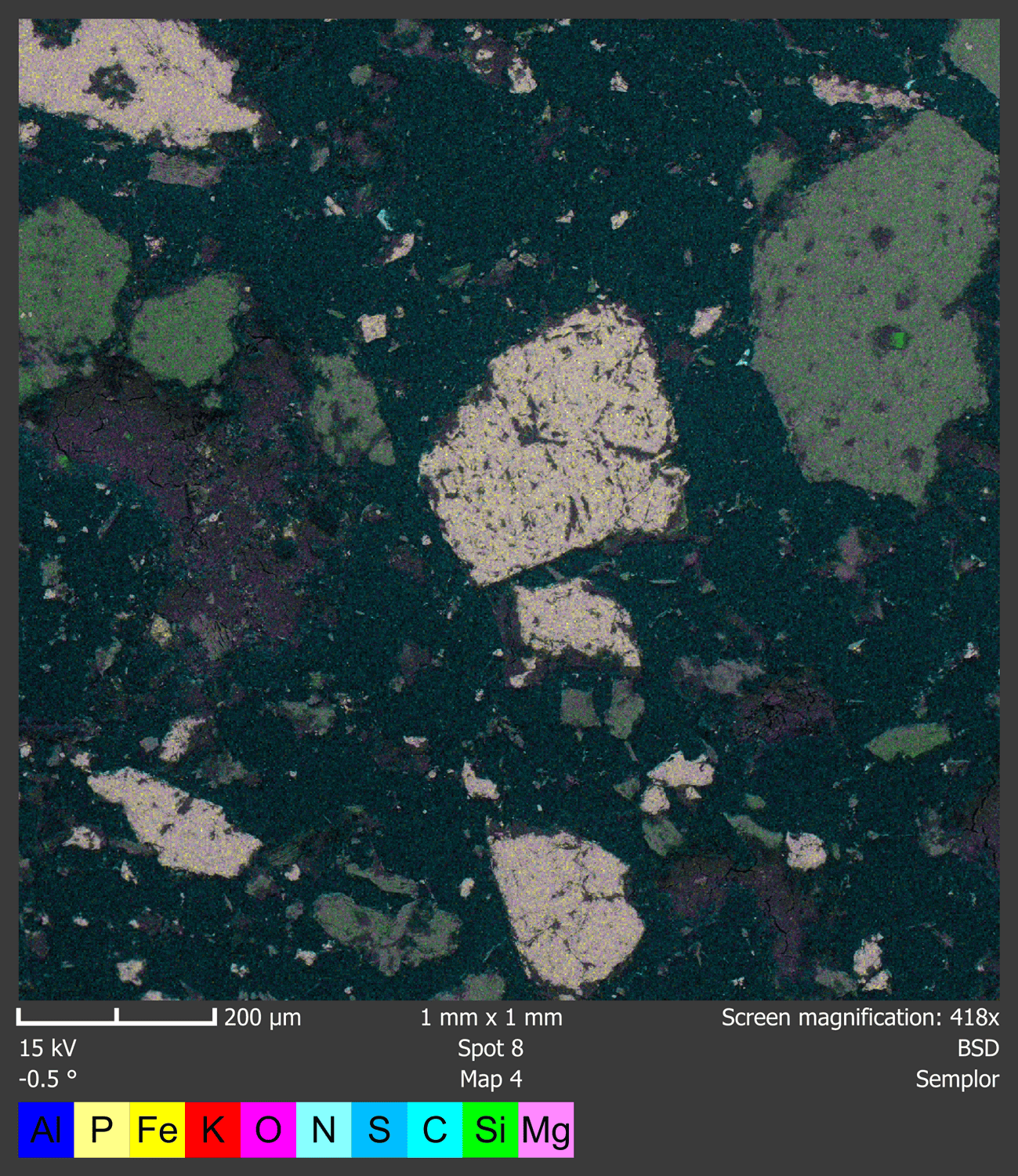
Semplor EDS elemental map of the same iron-ore specimen field

Fig 02 · Elemental map of the same field

Purpose & when to use

Scanning electron microscopy with energy-dispersive X-ray spectroscopy (SEM-EDS) links a particle's appearance to the elements detected at a selected point, along a line, or across a mapped area. High-magnification images document projected size, shape, surface texture, fractures, fibers, agglomerates, and inclusions. EDS adds elemental evidence to help narrow a source hypothesis, distinguish unlike materials, or guide the next analytical step. Use the service when:

  1. Batch-contamination or complaint investigations compare an unknown particle with raw materials, packaging, and process residues to support FDA regulated root-cause documentation.
  2. Inhalation powder teams compare particle form before and after blending, micronization, filling, or device actuation within an FDA CMC development program.
  3. Medical-device teams examine wear debris, coating fragments, and foreign material using ASTM F1877 terminology where it fits the investigation.
  4. Filter and data-center teams compare captured dust across locations, operating states, or suspected sources with ISO 17025 quality-system records.
  5. Coatings, electronics, and materials teams locate inclusions, residues, fractures, and elemental contrasts during ASTM E1508 informed failure analysis.

SEM-EDS works best when the question is specific and the analyst can compare representative particles, regions, or known references. EDS reports detected elements; it does not by itself establish a molecule, polymer, biological species, crystal structure, or definitive source. We explain these limits in the report and recommend follow-up chemistry when needed.

Built for investigations across products and materials

The same image-plus-elements workflow can answer different questions when sampling, preparation, and comparison rules are matched to the material and decision.

  • Pharma powdersSize, form, agglomerates, foreign matter
  • Medical devicesResidue, wear, coatings, inclusions
  • FiltersCaptured particles and deposits
  • ElectronicsDust, debris, process contamination
  • CoatingsSurface defects and elemental contrast

Instrumentation & measurement basis

A Semplor NANOS desktop SEM combines surface imaging, compositional contrast, and embedded EDS so we can examine morphology and elemental evidence together.

100 – 200000 xmagnification

Semplor NANOS SEM-EDS

Secondary-electron imaging resolves surface topography; four-quadrant backscattered-electron imaging adds compositional contrast. The embedded 30 mm² silicon-drift EDS detector supports point analysis, line analysis, and elemental mapping. The manufacturer's <8 nm resolution is an imaging specification, not a guaranteed particle-size measurement limit.

Test method options

MethodStrengthsTradeoffAligned with
Particle size and shape (SEM image analysis)
  • Measures projected dimensions and agreed shape descriptors from SEM images acquired under a project-specific sampling plan.
  • Documents angularity, aspect ratio, surface texture, fractures, fibers, pores, and agglomeration for selected particles and fields.
Two-dimensional projections and selected fields do not automatically represent a bulk population or aerodynamic size distribution.—
Elemental screening (EDS point, line, and map)
  • Targets individual particles or regions for elemental spectra, line profiles, and distribution maps.
  • Reports method-appropriate composition together with the limits of the selected acquisition and calculation approach.
EDS does not establish molecular, polymer, biological, or crystallographic identity and has sample-dependent detection limits.—
Foreign-particle investigation (unknown versus reference)
  • Combines morphology and elemental fingerprints to test whether an unknown is consistent with supplied source materials.
  • Annotated images keep each spectrum or map tied to the particle and region actually examined.
A source conclusion may remain qualified when candidate materials overlap or representative reference samples are unavailable.—
Surface and particle examination (materials troubleshooting)
  • Locates cracks, inclusions, deposits, surface boundaries, or wear features before elemental analysis is targeted.
  • Pairs secondary- and backscattered-electron views to separate topographic features from compositional contrast.
Mounting, coating, sample geometry, or vacuum compatibility can constrain the area and condition that can be examined.—

Setup configurations

We start with the decision: identify a foreign particle, compare materials, characterize powder morphology, or examine a defect. We then define the specimen, reference materials, preparation, and fields of view. The protocol also specifies particles or regions to analyze, image scales, the EDS collection plan, and acceptance or comparison rules. Replicate count is defined during protocol development.

Sample type

Submit dry powders, filters, wipes, coupons, fragments, components, or collected particles with handling history and the suspected problem clearly identified.

Reference materials

Provide candidate source materials, known-good parts, control lots, or process residues when the question involves comparison or likely origin.

Preparation

Mounting, dispersion, conductive coating, and high- or low-vacuum imaging are selected to preserve the feature and control charging or contamination.

Sampling plan

Agree on fields, magnifications, particle-selection rules, dimensions, shape descriptors, EDS targets, and minimum usable counts before analysis begins.

Comparison rules

Define how image features and elemental patterns will be compared and what language the evidence can support in the final report.

Key data outputs & reporting

Each conclusion in the report is tied to the exact image, particle, region, spectrum, or elemental map examined. Depending on scope, we report projected dimensions with image scale bars, shape descriptors, representative morphology, elemental peaks, method-appropriate composition, and spatial distribution. Unknown-versus-reference projects include side-by-side evidence tables showing where observations agree, where they differ, and which questions remain unresolved. Technical teams can use that evidence for disposition, root-cause work, formulation decisions, or follow-up testing.

Primary outputs

  • SEM images with particle dimensions, scale bars, and annotated regions of interest
  • Projected size and shape measurements using agreed descriptors and particle-selection rules
  • EDS point spectra, line profiles, and elemental maps tied to the analyzed image location
  • Qualitative, semi-quantitative, or quantitative elemental results with the calculation basis stated
  • Unknown-versus-reference comparison table with supported interpretations and unresolved alternatives

Deliverables

#FormatContents
01Technical reportMethods, preparation, sampling logic, images, EDS results, interpretation, and limitations.
02Image packageLabeled SEM images, spectra, line profiles, and elemental maps selected for the study.
03Data tablesParticle measurements and elemental results in CSV or XLSX format when included in scope.

QA / QC & data integrity

Controls depend on the question and sample. A foreign-particle investigation may use blank media, known-good product, and candidate source materials; a powder study may use documented field selection and repeat measurements. Instrument checks, preparation records, chain of custody, retained raw images and spectra, and reviewed calculations keep the findings traceable from specimen receipt through interpretation.

Blank stubs, media blanks, known-good samples, or reference materials are included where they can distinguish background and handling artifacts.

Magnification, detector, accelerating voltage, vacuum mode, working conditions, and image scale are recorded for each selected field.

EDS checks, acquisition settings, count statistics, peak assignments, corrections, and calculation basis remain with the raw spectral record.

Chain of custody connects sample identity, condition, preparation, mount location, image fields, EDS targets, and report figures.

Deviations, exclusions, particle-selection rules, acceptance criteria, and reviewer decisions are documented in the technical report.

Why ARE Labs

ARE Labs brings aerosol, inhalation, filtration, deposition, and device-testing experience to particle investigations. We design the imaging and elemental-analysis plan around your product decision, with controls and interpretation limits agreed at the start. Our lab's ISO/IEC 17025 accreditation is scope-limited; this new SEM-EDS service is not offered as an accredited test.

Reviewed byJamie Balarashti (25 yrs - cascade & inhalation methods) - Weston Schaper (7 yrs - real-time sizing & nanoparticle work)
900+Studies performed
300+Clients supported
17years (since March 2009)
17025Lab ISO quality system

Common questions

Teams usually arrive with an unknown particle, a material comparison, or a powder that behaves differently than expected. These answers explain how we select particles and imaging conditions, what EDS can establish, and what we need to scope the work. The final protocol depends on sample form, heterogeneity, reference availability, and the decision the results must support.

Q.Can SEM-EDS identify an unknown particle?
A.SEM-EDS documents morphology and detected elements so we can compare that evidence with candidate materials or references. The results may support a likely material class or source hypothesis, but EDS alone does not prove a molecule, polymer, biological species, or exact source.
Q.Can you measure particle size and shape?
A.Yes. We can measure projected dimensions and agreed two-dimensional shape descriptors from SEM images with scale bars. The sampling plan must define how fields and particles are selected. These measurements are not an aerodynamic particle-size distribution and do not automatically represent the bulk population.
Q.What samples should we send?
A.Send the unknown or test sample in a container or on media that protects the region of interest. Include known-good samples, suspected source materials, filters, blanks, or control lots when comparison is part of the question.
Q.What affects scope and turnaround?
A.Scope depends on sample count, preparation, vacuum compatibility, coating or sectioning needs, the number of fields and particles, EDS targets, references, and reporting depth. We agree on those choices before analysis so the quote reflects the work needed to support your decision.
Q.What data will we receive?
A.Deliverables can include calibrated SEM images, particle measurements, annotated regions, EDS spectra, line profiles, elemental maps, composition tables, and a technical report. Raw image and tabular exports can be included when requested during scoping.
Q.Does this replace chemical identification?
A.No. SEM-EDS provides morphology and elemental information. Molecular, polymer, phase, crystal, or biological identification may require FTIR, Raman, XRD, chromatography, mass spectrometry, or microbiological methods selected for the sample and question.

Standards & guidance

Testing can be aligned with the references that fit the analytical endpoint. ISO 19749 frames SEM-based particle size and shape measurements, while ASTM F1877 addresses medical-device particle morphology. ISO 22309 and ASTM E1508 guide EDS quantitation and interpretation. A fit-for-purpose protocol defines sampling, preparation, fields, particles, calculations, controls, and reporting when no single standard governs the complete investigation.